Yolanda Sonnabend (1935-2015)

15Aug10_011_(640x480)
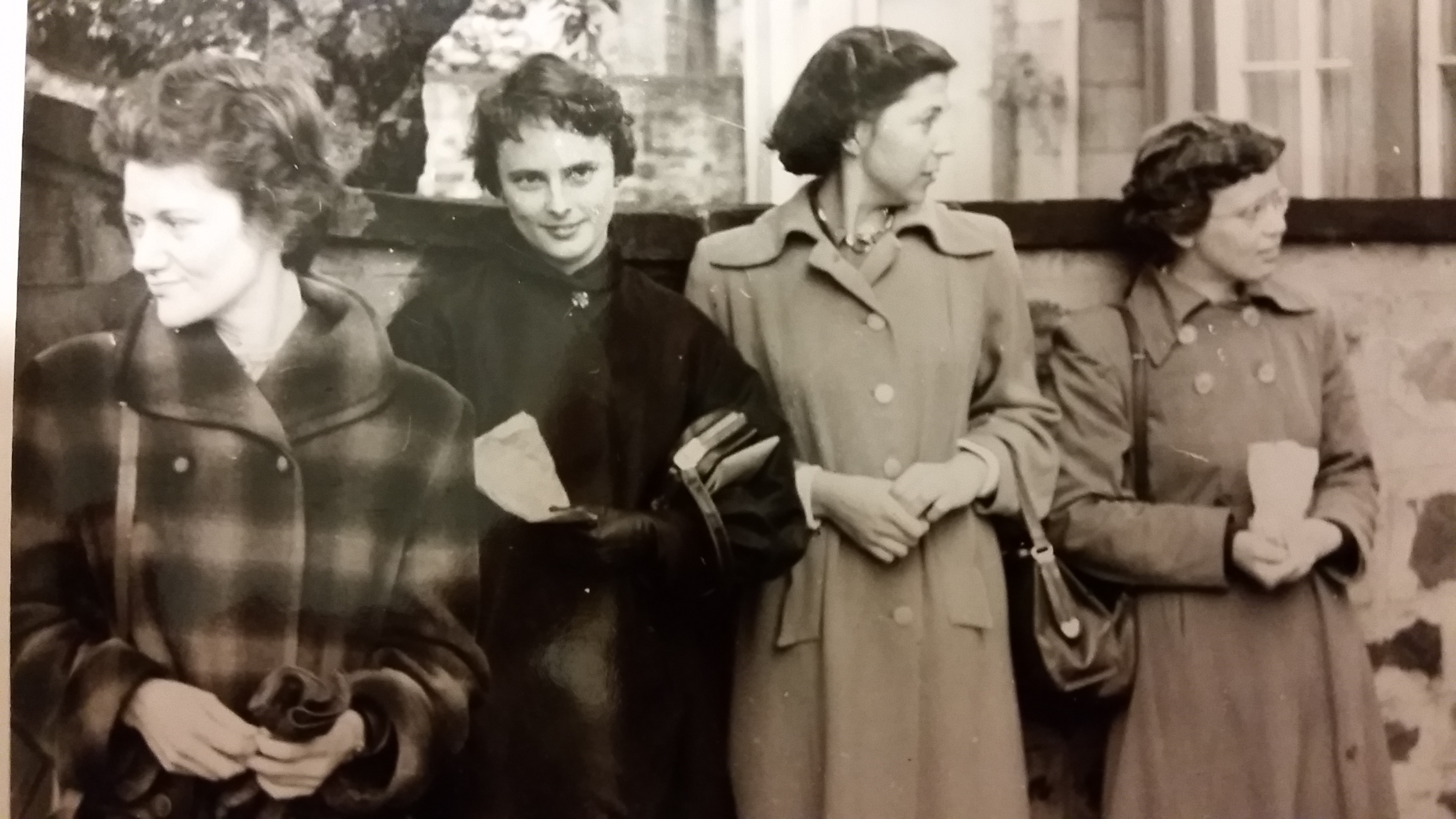
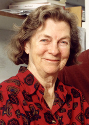
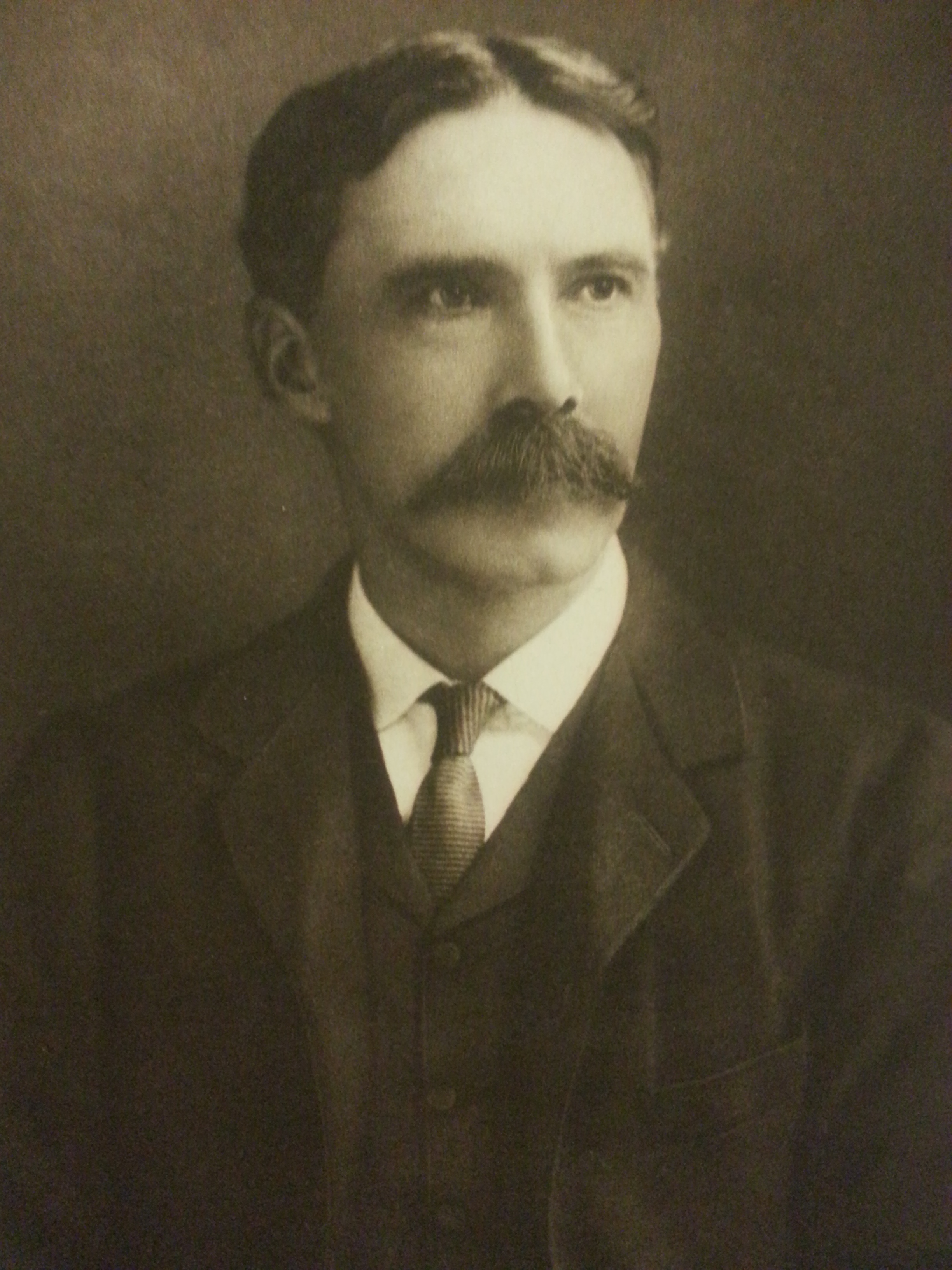
We were saddened to learn recently of the death of the artist and stage designer Yolanda Sonnabend, on 9 November 2015. In May 2013 we were delighted to acquire a collection of 200 or so artworks by Sonnabend, produced in collaboration with the developmental biologist C.H. Waddington, whose papers were catalogued as part of the ‘Towards Dolly’ project. This collection was first viewed in 2010 by our colleague Graeme Eddie, who visited Sonnabend in her home and took the photographs featured here.
15Aug10_024_(640x480)Yolanda Pauline Tamara Sonnabend was born on 26 March 1935 in Bulawayo, Southern
Rhodesia (now Zimbabwe) to German-Russian parents. After a cultured upbringing, Yolanda went on to study art in Geneva, and painting and stage design at the Slade School of Fine Art, London. She received her first design commission at the Royal Ballet in 1958, while she was still an undergraduate, and went on to forge a brilliant career as a stage designer. She worked closely with the choreographer Sir Kenneth MacMillan throughout the 1960s-1980s, and designed for theatre, opera and ballet shows across Europe, as well as designing Derek Jarman’s 1979 film adaptation of The Tempest. She was also an acclaimed portrait painter, whose subjects included Stephen Hawking and Steven Berkoff. In 2000 Sonnabend was awarded the Garrick/Milne Prize for theatrical portraiture.15Aug10_025_(640x480)

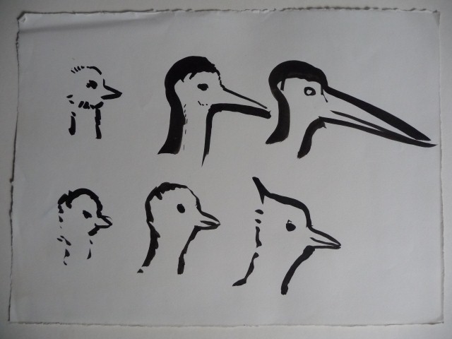
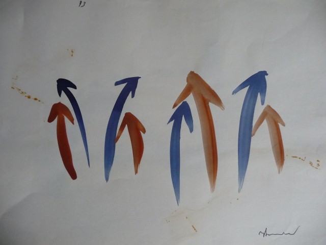
Sonnabend’s friendship and collaboration with C.H. Waddington is captured in the illustrations she produced for his book Tools for Thought: how to understand and apply the latest scientific techniques of problem solving. Waddington was a biologist and embryologist, and at the time he knew Sonnabend, was the director of the Institute of Animal Genetics in Edinburgh. However, Waddington’s interests extended far beyond the purely scientific, encompassing art, architecture, ecology, robotics and early computing; he was also committed to communicating with the wider public about these topics. Tools for Thought was his final book, published posthumously in 1977, and was intended to be a popular guide to new ways of perceiving and understanding the world’s scientific, political and ecological problems. Sonnabend’s stark and imaginative pen and ink drawings formed the perfect ‘other half’ to the book; incorporating triangles, graphs, arrows and bird heads.15Aug10_022_(640x480)

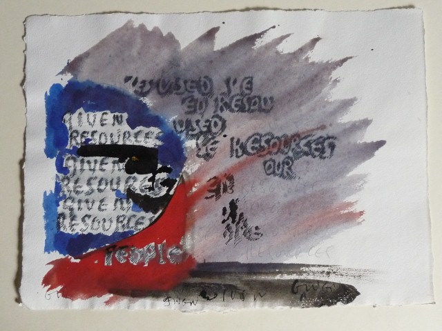
Many of Sonnabend’s designs were, sadly, not included in the final book (Waddington died during the proofing stage), but the originals exist in the collection acquired by the Library. 15Aug10_017_(480x640)
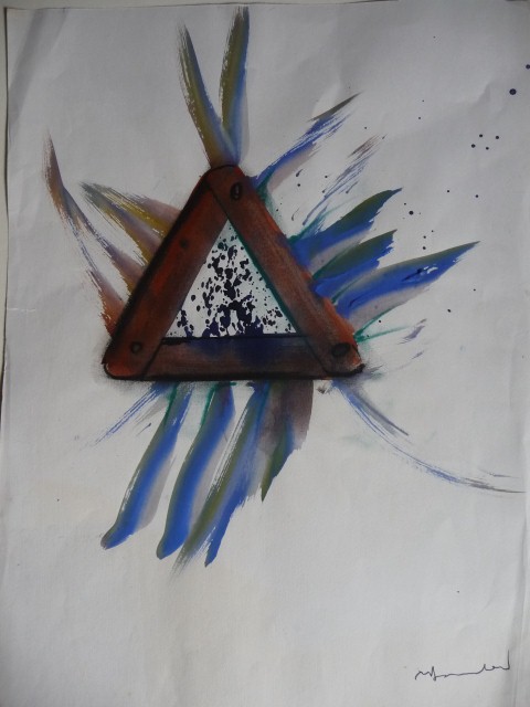
Sketches on graph paper and collages made with shredded magazine articles sit alongside completed, signed pieces. The correspondence which accompanies these works shows the affectionate and intellectual stimulating relationship Sonnabend and Waddington shared.

Sonnabend’s artistic legacy lives on in the portraits collected by the National Portrait Gallery and the Science Museum and the Design Collection at the Royal Opera House – but her work for C.H. Waddington sheds a new light on both their careers.

With thanks to Graeme Eddie and Dr Joseph Sonnabend.

Clare Button
Project Archivist

Dolly on Display

Collection: University of Edinburgh; Persons: ; Event: Towards Dolly; Place: Main Library; The University of Edinburgh; Category: Science; Genetics; University Exhibition; Description: The opening night of the Towards Dolly exhibition in the exhibition room on the ground floor of the Main Library, 30th July 2015.

All images courtesy of the Digital Imaging Unit

12 days ago, the doors of Edinburgh University’s Main Library Exhibition Gallery opened on ‘Towards Dolly: a century of animal genetics in Edinburgh’. This exhibition has given us the chance to display Dolly the sheep, on loan from National Museums Scotland, while her new home in their redesigned Science and Technology Gallery is being prepared. But it has also allowed us to show some of the collections which have been catalogued over the last three years as part of the Wellcome Trust-funded ‘Towards Dolly’, ‘Making of Dolly’ and ‘Science on a Plate’ projects, which tell the story of animal genetics in Edinburgh over the last 100 years. We’ve also been able to feature the microscope which cloned Dolly (on loan from the Roslin Institute), DNA models on loan from the School of Biological Sciences, books from the Noreen and Kenneth Murray Library and the Main Library’s general holdings, and animal skulls from the University’s Anatomical Museum. Together, these varied items represent the richness of the University’s collections, the diversity of Edinburgh’s genetics history, and the generosity of our colleagues in helping us source the perfect exhibits.

0068006dWorking together with our designers Studio SP and CRC Exhibitions Officer Emma Smith, we decided to take a chronological approach, beginning with James Cossar Ewart’s early zebra and horse cross-breeding work in Penicuik and ending by looking at cutting-edge stem research taking place in the University’s MRC Centre for Regenerative Medicine. We aimed to represent just some of the many animal breeding and genetics organisations which have existed and intersected over the decades: the Institute of Animal Genetics, the Animal Breeding Research Organisation, the Poultry Research Centre, the Institute of Animal Physiology and Genetics Research and the Roslin Institute. We also highlight outstanding work by particular individuals, such as Charlotte Auerbach’s work on mustard gas with J.M. Robson during World War Two and Douglas Falconer’s landmark textbook Introduction to Quantitative Genetics.

Collection: University of Edinburgh; Persons: ; Event: Towards Dolly; Place: Main Library; The University of Edinburgh; Category: Science; Genetics; University Exhibition; Description: The opening night of the Towards Dolly exhibition in the exhibition room on the ground floor of the Main Library, 30th July 2015.

Dolly stands proudly in her own case, and already staff and members of the public are enjoying getting up close to her eyeline and even sharing their ‘Dolly selfies’ on social media! An informational panel behind Dolly’s case tells the story of the other groundbreaking sheep at Roslin: from Tracy the transgenic sheep, who contained a human gene for a therapeutic protein to potentially treat lung disorders, to Megan and Morag, the twin sheep who made Dolly possible, to Polly the sheep and her siblings, who were cloned and transgenic at the same time.

Collection: University of Edinburgh; Persons: ; Event: Towards Dolly; Place: Main Library; The University of Edinburgh; Category: Science; Genetics; University Exhibition; Description: The opening night of the Towards Dolly exhibition in the exhibition room on the ground floor of the Main Library, 30th July 2015.

As well as physical exhibits, there is pioneering filmmaker Eric Lucey’s film of the developing egg of the fruit fly Drosophila – this fly has played a hugely important role in the history of genetics, as we also explore in a case in the external entrance area to the exhibition (pictured above). In addition, audio clips of scientists talking about their work are available for visitors to hear on their smartphones via the IZI Travel app or any QR code reader; from Ian Wilmut recalling the reaction of the public, media and government to Dolly’s birth to Institute of Animal Genetics staff singing comedy genetics songs!

Collection: University of Edinburgh; Persons: ; Event: Towards Dolly; Place: Main Library; The University of Edinburgh; Category: Science; Genetics; University Exhibition; Description: The opening night of the Towards Dolly exhibition in the exhibition room on the ground floor of the Main Library, 30th July 2015.

It has been good to hear positive responses to the exhibition from current and retired scientists as well as the family of many individuals we have featured. The exhibition was recently selected as one of ThreeWeeks magazine’s ’81 recommended shows’ to see during the Edinburgh Festival. If you haven’t already, do come along, take a look, and tell us what you think!

logo_only

‘Towards Dolly: a century of animal genetics in Edinburgh’ is free and open to the public 10:00-17.00 Monday to Saturday, at the Main Library Exhibition Gallery, George Square, from 31 July-31 October 2015.
https://exhibitions.ed.ac.uk/towardsdolly
#towardsdolly

Clare Button
Project Archivist


A Visit from Dolly…

It has been a while since my last post, but rest assured, the ‘Towards Dolly’ project has not been idle! As you’ll have seen from the last couple of posts, John Bryden has done a brilliant job digitising the Roslin glass slide collection. We’ve also been conducting some fascinating oral history recordings which we’ll post more about soon. But mainly, we’re excited to announce our forthcoming exhibition in the University of Edinburgh Main Library’s Exhibition Gallery. Titled ‘Towards Dolly: a century of animal genetics in Edinburgh’, this multimedia exhibition will explore the enormous contribution the Edinburgh area has made to the science of genetics over the last century. We will be open from 31 July to 31 October 2015 and also appear as part of this year’s Edinburgh Festival Fringe Festival: https://tickets.edfringe.com/whats-on/towards-dolly-a-century-of-animal-genetics-in-edinburgh

Continue reading

Digitisation of the Roslin Glass Slide Collection; complete!

That’s me signing out, 3460 glass plate slides later. It is hard to know where to start. Lots of images of cows, pigs, horses and sheep! I now have the satisfaction of viewing the entire collection on our online digital collections platform – Roslin Slides. Furthermore, I was glad to find out that a selection of the slides will be shown at the Towards Dolly exhibition, here on the ground floor of the main library, in July 2015. With so many rich and diverse images, the best place for them is out in the open, easily accessible to academics and members of the public. This one of the many benefits of digital collections. The images are there to be enjoyed and engaged with; an activity that is not so easy when slides are neatly stacked away on shelves in a strong room! With the latter in mind, here are just a few more of my favourite slides from the collection.

Silver Spangled Hamburghs

Silver Spangled Hamburghs

Prize Cock and Hen Ostriches, South Africa

Prize Cock and Hen Ostriches, South Africa

Professor Robert Wallace

Professor Robert Wallace

'Hiawatha', Clydesdale horse

‘Hiawatha’, Clydesdale horse

Perth Ram Sales

Perth Ram Sales

Spy Pig

Spy Pig

Jersey Collings Cow

Jersey Collings Cow

Khonds in Phulbani, Khandmal, Orissa, India

Khonds in Phulbani, Khandmal, Orissa, India

The 'Wandsworth Lion'

The ‘Wandsworth Lion’

Zebra

Zebra

John

Project Photographer

‘Science on a Plate’

Science on a Plate: an evolution

I have been based in the Digital Imaging Unit, digitising the Roslin Glass Plate Slide Collection, since October last year as part of the ‘Science on a Plate’ project, funded by the Wellcome Trust’s Research Resources scheme. Recently, I became intrigued by the history of the glass plate slide itself so I decided to carry out a little research of my own.

Curious to discover where glass plate slides are positioned on the timeline of photography; what chemistry and techniques were involved in their making; and the role they played, I began with searching online and delving into some books on the history of photography.

The slides in the Roslin Collection date, loosely, between the late nineteenth century and the early twentieth century. This leads me to think that the majority of the slides are dry plate, gelatin-coated, slides as this was the most popular process used during that time period. Other processes, however, may have also crept in to the collection because different techniques and methods may have overlapped.

The slides are all positive, transparent images, with the nature of the images spanning; documentary photography, maps, statistics, instructions, illustrations, portraits, images taken from publications… the list goes on. Physically, the slides are all 3×3 inches in width and diameter, with a depth of roughly 3-6 millimetres. There are 3465 of them!

To give a little context, these slides might also be referred to as lantern slides. This is a term that relates to the fact that, early on, magic lanterns would be used to project similar looking slides. Magic Lantern (and Sciopticon) projectors had been used long before the invention of photography to project images painted on glass.

0053658d

Roslin glass slide (or lantern slide). Illustration of two llamas standing on a rocky ledge.

It was not until 1840 that the inventors of the daguerreotype began using magic lantern projectors in an attempt to project their photographic images for better viewing. The dull and foggy nature of the daguerreotype, however, did not lend itself well to the projection of light.

Next came the creation of the wet plate negative in 1851 by the Englishman, Frederick Scott Archer. This process involved coating glass plates with a collodian chemical and exposing light onto the glass while the chemical was still wet. This allowed for shorter exposure times when taking photographs. Previously, daguerreotypes (1839-1850’s) and calotypes (1841-1850’s) would require long exposures, often over one minute, forcing those being photographed to remain still in order to avoid blurring within the image. This is why daguerreotypes and calotypes often look posed and overly considered as compositions.

Newhaven 14. 7 calotypes, 6 carbons, print size 4.

Calotype by Hill and Adamson, 1840’s

Accordingly, the colllodian wet plates gave a bit more flexibility when composing photographs. If, however, using the wet glass plate outwith the studio, photographers would have been required to carry processing chemicals and a portable darkroom as the wet plates would need developing immediately after exposure.

Photography remained a rather laborious procedure until the invention of the dry plate slide. In 1871, Richard Leach Maddox discovered that coating glass with silver bromide within a layer of gelatin would give more efficient photographic results; speedier and with a longer shelf life. The burden of hauling around chemicals and a darkroom was removed because the developing could be done at a later date by a specialist. In light of this photography became more accessible, opening up the practice to amateur photographers. Only the camera (camera models were becoming smaller and more portable over time) and the dry plates themselves were needed. Dry glass plates could be produced on mass, ready to use on the job. The act of taking a photograph therefore became less time-consuming, causing a change in the style of photographs being produced. This can be seen in the Roslin Glass Plate Slide Collection. On viewing many of the slides the camera seems to be there as a spectator, very much in the moment, giving the viewer a sense of what it mean to ‘be there’ – a relatively new phenomenon.

0053978d

Roslin glass slide. A street hawker with his horse drawn cart standing next to a couple of children in Buenos Aires, Argentina in the late 19th or early 20th century.

0053041d

Roslin glass slide. A group of men eating breakfast in their camp on the plains in [Argentina or Uruguay] in the early 20th century.

0053159d

Roslin glass slide. Men using a machine to press hay in [South Africa] in the early 20th century.

Throughout the second half of the nineteenth century, photography had become recognised as the best tool for recording reality; the world around us. Whilst some were fervently trying to establish photography as an artistic medium in its own right, others were more concerned with employing the camera as a way of recording the scenes before them. The latter approach can be seen throughout the Roslin slides as new territories are explored and recorded.

Of all the various glass plate processes, the dry, gelatin-based plate had the longest run of success. It was not until the 1930’s when it was reduced to history with the introduction of nitrate film negatives and, namely the 35mm Kodachrome.

These slides were catalogued as part of the ‘Towards Dolly’ project and the Roslin Slide Collection can be viewed here.

Some useful reading:

Frizot, Michel, The New History of Photography (1994), Chapter 5

http://memory.loc.gov/ammem/collections/landscape/lanternhistory.html

http://archives.syr.edu/exhibits/glassplate_about.html

http://www.nfsa.gov.au/research/papers/2012/05/10/preserving-20th-century-glass-cinema-slides/


John

Project Photographer: Science on a Plate.

Mary Lyon (1925-2014) – Edinburgh connections

M.M. Perry, R. Phillips, S. Dare-Delaney, M. Lyon, Edinburgh (1950s), from EUA IN1/ACU/A1/5/7

M.M. Perry, R. Phillips, S. Dare-Delaney, M. Lyon, Edinburgh (1950s), from EUA IN1/ACU/A1/5/7

We were saddened to hear recently of the passing of Mary Lyon, a distinguished mouse geneticist. Born in 1925 in Norwich, Mary was best known for her X-chromosome inactivation hypothesis, which proposed that one of the two X chromosomes in every cell of female mammals is inactivated. Mary worked at the MRC Radiobiology Unit in Harwell from 1955 until her death, becoming head of the genetics division (later the Mammalian Genetics Unit) in 1995. What is perhaps not so well-known is that her early work took place in Edinburgh, at the Institute of Animal Genetics.

Mary began at the Institute in 1948 to continue her PhD on mouse genetics, which she had begun in Cambridge under R.A. Fisher. This was after studying zoology at Girton College, Cambridge (although, as women were not allowed to be official members of the University until 1948, Mary was only awarded a ‘titular degree’). The Institute of Animal Genetics, then under the directorship of C.H. Waddington, possessed superior histology facilities, which she needed for her work. Mary ended up staying for a further five years after her PhD, working with Toby Carter on a project funded by the Medical Research Council to study mutagenesis in mice (this was at a time, following the Second World War and atomic bombs in Japan, of great concerns about the effects of nucelar fallout in the atmosphere). In a 2010 interview, Mary Lyon stated that, out of her whole career, it was her time in Edinburgh that she enjoyed the most: ‘It was a very lively academic atmosphere…a big genetics lab and a lot of able and enthusiastic geneticists.’ The above photograph, from the Institute of Animal Genetics archives, shows Mary (far right) with (right to left) Institute Librarian Stella Dare-Delaney, Mary’s assistant Rita Phillips, and distinguished molecular geneticist and embryologist Margaret Perry.

Toby Carter’s Mutagenesis Unit moved south to Harwell in order to find more space in which to breed and keep mice, taking Mary with it, as well as Rita Phillips. Scientists working with Douglas Falconer in Edinburgh had been the first to discover X-linked mutants in mice. With this discovery in mind, Mary, noticed that female mice carrying X-linked coat colour mutations had mottled coats. Male mice which inherited a mottled coat (i.e. a mutant gene on their single X-chromosome) all died, but the females survived. This must mean that the female possessed one, inactivated, mutant gene on one X-chromosome, but a normal gene on the other chromosome, which was activated – therefore a female mouse needs only one X chromosome for normal development. This inactivation of one of the two X chromosomes in the cells of females is still called ‘Lyonisation’, and the discovery had profound implications for understanding the genetic basis of X-linked diseases such as Duchenne Muscular Dystrophy. Grahame Bulfield, later director of the Roslin Institute, first positioned the mouse muscular dystrophy mutant on the X-chromosome using Mary’s stock of mouse X-chromosome mutants.

Over the next six decades, Mary also made important studies of Chromosome 17 and ‘the t-complex’, which had significant bearings on the understanding of non-Mendelian inheritance (a departure from the expected one-to-one ratio due to the abnormal segregation of chromosome pairs). Mary’s work pioneered the use of the mouse as a model organism for advances in cell and developmental biology as well as molecular medicine, and laid the foundations for comprehending the human genome. She chaired the Committee on Standardised Genetic Nomenclature for Mice from 1975 to 1990, was made a foreign associate of the US National Academy of Sciences, and was a Fellow of the Royal Society (being the 28th woman to be elected such). In 2004, the Mary Lyon Centre opened at Harwell, a leading international centre for mouse genetics, and in 2014 the UK Genetics Society created the Mary Lyon medal.

Mary died on Christmas Day 2014, aged 89, ‘after drinking a glass of sherry, eating
her Christmas lunch and settling down in her favourite chair for a nap’.

The University of Edinburgh’s remembrance of Mary Lyon can be read here: http://www.ed.ac.uk/news/staff/obituaries/2015/mary-lyon-030215

Clare Button
Project Archivist

Sources:

– ‘The Gift of Observation: An Interview with Mary Lyon’, Jane Gitschier (2010), http://journals.plos.org/plosgenetics/article?id=10.1371/journal.pgen.1000813
– ‘Mary F. Lyon (1925-2014): Grande dame of mouse genetics’, Sohaila Rastan, Nature, 518, (05 February 2015)

Frozen: The story of Frostie the calf

Frostie calf CROPIan Wilmut is best known for his involvement with the team which cloned Dolly the sheep in 1996. However, his scientific career, which spans more than five decades, includes a variety of groundbreaking achievements and discoveries, which are being revealed as his papers are catalogued as part of the continuing ‘Towards Dolly’ and ‘The Making of Dolly’ projects.

Ian Wilmut initially wished to work as a farmer, but he ‘shuffled sideways into scientific research’*, as he puts it, and ultimately won a scholarship as an undergraduate at Nottingham University to work for two months under Chris Polge at  the Animal Research Station in Cambridge. His role was to help out generally with experiments, but he soon became fascinated by embryos, and returned to work with Polge once he graduated. Wilmut’s PhD, awarded in 1971, was on the freezing of boar semen. Copies of Wilmut’s published papers exist in the archive from 1969 onwards, and a glance through the papers which appeared over the next decade reveal Wilmut’s wide-ranging research on the effects of freezing, thawing and warming on embryos and spermatozoa in mice, sheep and cattle.

In 1973, Wilmut was the first scientist to successfully freeze a calf embryo (using liquid nitrogen), thaw it, and transfer it to a surrogate mother. This process led to the birth of a healthy red and white Hereford-Friesian cross calf, which Wilmut wryly named ‘Frostie’. Wilmut’s findings were published within a few weeks of Frostie’s birth in The Veterinary Record as ‘Experiments on the low-temperature preservation of cow embryos’ (June 30 1973, 686-690), and a copy of the reprint survives in the archives. Wilmut has remarked that this was ‘one of the fastest scientific publications ever’. It also led to some considerable interest from the world’s media, with Wilmut appearing on television and newspapers from as far away as New Zealand seizing upon the story. This media attention was a precursor to the storm which Wilmut, Keith Campbell and team would generate two decades later when Dolly the sheep was born. Wilmut’s discovery of the viability of frozen embryos to produce healthy offspring has since been used across many different species in agriculture and also for the conservation of rare breeds. The first human to be born from a frozen embryo was Zoe Leyland, born in Melbourne in 1984.

Wilmut’s work with Chris Polge equipped him with many of the techniques in reproductive physiology which would instruct his later work on cloning, nuclear transfer, stem cell and regenerative medicine in Edinburgh, where Wilmut moved in 1973. Throughout his career Wilmut has been inspired by the possibilities of advances in reproductive physiology and biotechnology for fertility treatments,practical applications to the farming industry and breakthroughs in treatments or cures for debilitating genetic diseases.

*All quotes taken from The Second Creation: the age of biological control by the scientists who cloned Dolly, I. Wilmut. K. Campbell and C. Tudge (London, 2000).
Clare Button
Project Archivist

Science From The Tomb…

Coll-1364/1/6

Coll-1364/1/6: Papers of R.A. Beatty

It’s nearly Hallowe’en, when spooky subjects are foremost in our minds. An ideal time, then, to look at some rather unusual correspondence from the Richard Alan Beatty archive about Egyptian mummies! At first glance, this might seem an unlikely research subject for a reproductive physiologist, but Beatty had his reasons. Writing from the Institute of Animal Genetics to the Department of Egyptian Antiquities at the British Museum in July 1977, Beatty asks whether he may have a sample of ‘a testis of an Egyptian mummy’ to enable him to assess whether ‘ certain aspects of chromosome structure and spermatozoan morphology are stable’. In his letter, Beatty realises his request may be a ‘long shot’, but if it worked, ‘it could make an entertaining letter to Nature.’

Beatty was to be disappointed at first. He received a reply three days later from the Keeper of Egyptian Antiquities at the British Museum regretting that, as all their mummies were still in their wrapped state, the Museum could not allow any ‘surgical operation’ to take place. In reply, Beatty understands this restriction, but wonders if he could obtain any mummified cats instead, as ‘there would be merit in looking first at a mummy of some mammal other than man.’ He adds: ‘I read that 100,000 mummified cats were sold for fertiliser in the last century, and this made me hope that cats are in plentiful supply!’ However, he learned that those mummified animals in the Department’s collection were wrapped as well, and so also unavailable for study.

However, Beatty was directed to the Museum’s Department of Zoology, where he had better luck. This Department boasted a collection of mummified ‘monkeys, cats, dogs, and mongooses’, and were happy to let Beatty take a testis sample from an adult male dog from the W.M. Flinders Petrie collection, which was in an unwrapped state. He would also be permitted a sample from a human mummy in the Department of Palaeontology. Beatty visited the Museum on 16 December 1977 to take his samples, having been advised that ‘a strong sharp scalpel’ would be needed, the consistency of the mummified tissue being like ‘very hard leather’. Ever prepared, Beatty tested out his scalpel on ‘an old leather boot’ beforehand!

From a report amidst the correspondence, it appears Beatty was eventually successful in getting his samples from the dog and human mummies:

Testis cores taken 16/12/77, wrapped in polythene, placed in tube, tube later maintained in dessicator.

Dog: Consistency very hard – almost rock-like…

Human: Consistency like medium hard cheese, very oily in texture.

It is not clear from Beatty’s archive exactly what resulted from his research on the Egyptian mummies – so we’d be delighted to hear from anyone who may know more about it! In the meantime, you can read more about the strange story, mentioned by Beatty, of the 180,000 mummified cats brought over to England from Egypt in the nineteenth century to be used as fertiliser here:
http://www.strangehistory.net/2013/12/18/tens-of-thousands-of-egyptian-mummies-in-english-soil/

Happy Hallowe’en everyone!

Clare Button
Project Archivist

Dame Anne McLaren (1927-2007)

Anne McLaren (picture sourced from http://en.wikipedia.org/wiki/Anne_McLaren)

Anne McLaren (picture sourced from http://en.wikipedia.org

It’s always great to receive comments and feedback from our blog readers, especially those which suggest subjects or people which we haven’t yet featured. We have a growing list of posts to respond to our readers’ suggestions, and we are delighted that our first of these focuses on Dame Anne McLaren.

Anne McLaren was a hugely important figure in the fields of mammalian reproductive and developmental biology and genetics, and she is possibly best known for her work as director of the Medical Research Council Mammalian Development Unit at University College London. Her long and rich career in the techniques and ethics of fertility is covered in ample detail in John Biggers’ excellent obituary: http://www.theguardian.com/science/2007/jul/10/uk.obituaries

In this blog, I want to focus specifically on McLaren’s time in Edinburgh, and her appearances in our archive collections. After gaining her degree at Oxford and completing postdoctoral work in London, McLaren moved to Edinburgh in 1959 with her then-husband Donald Michie. She joined the Agricultural Research Council’s Unit of Animal Genetics (based within the Institute of Animal Genetics), working initially on the reproductive physiology of the mouse with Alan Beatty and others. During her time at the Institute, McLaren’s research spanned mammalian fertility, embryo transfer techniques and immunocontraception. She was particularly interested in egg transfer, the hormonal control of ovulation, superovulation and its effects on pregnancy, placental and foetal growth, and the effects of the uterine environment on skeletal character. She and John Biggers were the first to demonstrate that a mammalian embryo grown in vitro for several days would subsequently develop into a normal adult. McLaren also worked with chimeras (organisms consisting of two or more genetically different kinds of tissue), and her later book on the subject, published in 1976, became a classic text.

McLaren was well liked and respected in Edinburgh. The correspondence of Institute of Animal Genetics director C.H. Waddington reveals that he proposed McLaren for Fellowship of the Royal Society in 1968 (he was disappointed that she was not elected until 1974, after she had left the Institute), with F.W. Rogers Brambell as seconder. Waddington’s statement of support commends McLaren’s work on the reproductive biology of the mouse, and in particular the rigour of her statistical and quantitative approach. Waddington also praises her personal attributes: ‘She has in a high degree an ability found only in some scientists of being both highly critical and extremely helpful. Very many workers, at all levels from the young to quite mature ones, like to talk over with her some subject they are tackling, confident that she will spot any weaknesses in their arguments, or, more positively, coax them into thinking straighter than they had done before.’ (Coll-41/9/4/4)

Alan Beatty’s papers contain the most information relating to McLaren, as they worked closely together until McLaren departed for London in 1974. They secured a series of grants from the Ford Foundation for a sustained programme of work on reproductive physiology, and together with colleagues, they lobbied for a Centre for Reproductive Biology to be established in Edinburgh (which occurred in 1980). Beatty’s archive reveals an active and busy schedule of planning meetings and funding applications underpinning he and McLaren’s research. Letters from McLaren after her departure to London show that she continued to take an interest in matters in Edinburgh, and stayed in touch with old colleagues and friends.

In London, McLaren’s research took her on to study the development of mammalian primordial germ cells, and she published an acclaimed book on the subject in 1980. After her retirement from the MGU, she became principal research associate at the Welcome Trust/Cancer Research UK Gurdon Institute in Cambridge. Anne McLaren died aged 80, along with her former husband Donald Michie, in a car accident en route from Cambridge to London on 7 July 2007.

Importantly, McLaren’s scientific work formed the basis of her wider engagement with ethical and societal issues surrounding fertility and reproduction, and, later, stem cell research and cloning technologies. She sat on the Warnock Committee, which contributed to the passing of the 1987 Family Law Reform Act and the 1990 Human Fertilisation and Embryology Act. A copy of one of her articles, titled ‘The Future of the Family’, was retained by Waddington in his papers, and demonstrates that, for McLaren, science has the potential to direct humans towards a better society. The article, written in 1972, concludes:

People in the future will be faced with more leisure: I hope that they will use it for a greater degree of democratic participation in the running of our society; for self-education, aided by computers whose tutorial intelligences will soon be at our disposal; and above all for the benefit of their smaller families of children. It is in the early years of life that personalities are moulded and the foundations of ethical systems laid: we must find out how not to cripple children’s minds as most, perhaps all, are crippled today, and how to instil an ethic of loyalty and protectiveness which begins with other members of the family and does not stop at national boundaries, but extends for the first time over the entire human race.

Clare Button
Project Archivist
If you have a topic relating to animal genetics in Edinburgh which you’d like to see covered, get in touch!


Freshers’ Week – 1870 style

Early portrait of James Cossar Ewart (GB 237 Coll-14/4/2)

Early portrait of James Cossar Ewart (GB 237 Coll-14/4/2)

This week is one of the busiest times of the year for the University of Edinburgh, and a momentous week for our brand new students – it’s Fresher’s Week!

The ‘Towards Dolly’ collections are rich in detail about the research and careers of Edinburgh’s scientists, but there isn’t so much concerning the early parts of their lives. So I was especially surprised when, cataloguing the archives of James Cossar Ewart (Professor of Natural History, 1882-1927) during an earlier part of the project, I came across a perfectly preserved diary from his undergraduate days. This small volume, filled with Ewart’s flowery but sometimes erratic handwriting, captures precious details from his own first experiences of student life. In some ways, they don’t differ hugely from the experiences of a fresher from today; in others, they reveal a University on the brink of major changes, particularly where the education of women was concerned. Continue reading