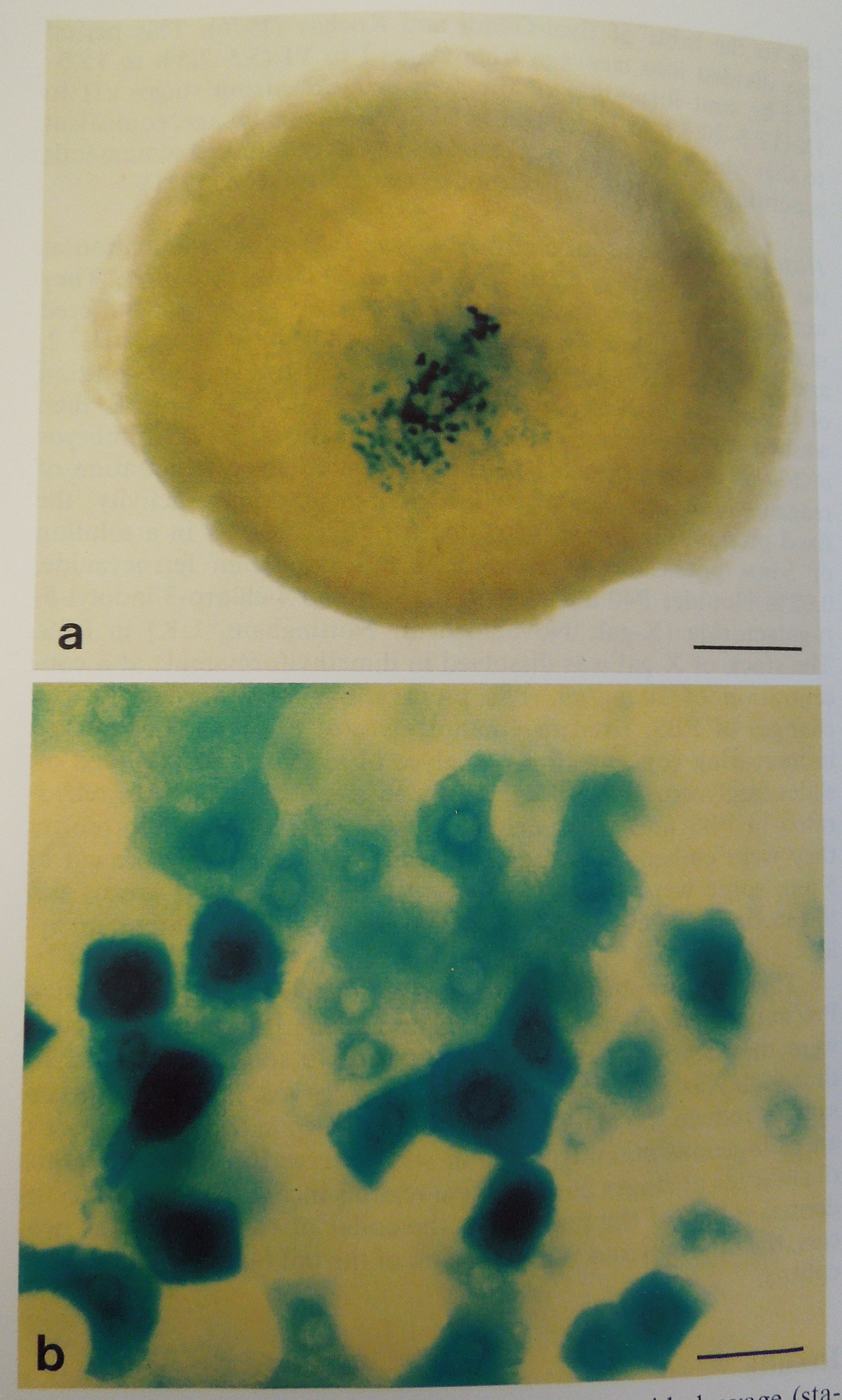
Margaret Perry, David Morrice and Helen Sang’s article, ‘Expression of exogenous DNA during the early development of the chick embryo’ in Roux’s Archive of Developmental Biology, Vol. 2, 1991, p. 302-319, discusses how they created a ‘plasmid construct containing the reporter gene, lacZ, under the control of the cytomegalovirus immediate early promoter, [which] was injected into the germinal disc of fertilised chick ova.’ The image, Fig. 2a, b, shows a ‘whole mount of a chick embryo at mid-cleavage (Stage IV) following injection of a lacZ gene construct (pHFBGCM) into the fertilised ovum, in vitro  culture and 5-bromo-4chloro-3-indolyl-beta-D-Galactoside (X-gal) staining for beta-galactosidase. Stained blastomeres are present in the centre of the blastodisc (a). They vary in size and intensity of staining, and some are stained in the perinuclear zone (b). According to the article, the ‘results provide supportive evidence for transcriptional activity during the cleavage stages of avian development. They also confirm previous findings on the loss of exogenous DNA during the early development of the chick.’
As you may be aware, cell staining is a technique used by scientists in order to better visualise cells and their components under the microscope, and this example demonstrates both aspects admirably. While scientifically it allows for a clearer understanding the stages of avian development, it also seems to have an artistic component as well!

Fascinating stuff!